Noninvasive Elastography May Help in Diagnosis of Gaucher Disease Patients
Written by |
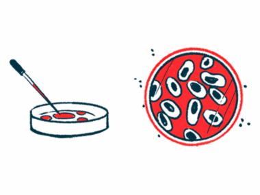
Noninvasive techniques measuring shear wave velocity in body tissues show potential at distinguishing Gaucher disease patients from other diseases with common symptoms, such as enlarged spleen.
The study, “Are transient and shear wave elastography useful tools in Gaucher disease?” was published in the journal Blood Cells, Molecules & Diseases.
Early tissue damage detection in Gaucher disease has been hampered by the lack of a reliable marker, either by imaging biochemical or imaging techniques.
In this study, authors investigated the potential for elastography as a strategy for evaluating patients with Gaucher disease. Transient elastography (TE) is a noninvasive method that works by measuring shear wave velocity through a certain tissue when subjected to different biomechanical properties. It was first developed to measure liver stiffness to quantify liver fibrosis. Because fibrotic tissues have increased stiffness, the shear wave propagates faster.
More recently, transient elastography has been used to measure spleen stiffness in patients with liver cirrhosis portal hypertension (high blood pressure in the hepatic portal system, which comprises the portal vein, the large vein that brings blood from the intestine to the liver, and its branches).
In this study, researchers determined liver and spleen stiffness in Gaucher disease patients. The study included 42 patients with Gaucher disease type 1, 33 patients with liver cirrhosis, and 22 healthy controls. They then applied transient and 2D shear wave elastography (SWE), a more recent modality where shear wave is integrated with an ultrasound device, thereby revealing both anatomy and tissue stiffness.
Researchers observed that using either applied transient or 2D shear wave elastography was a noninvasive method to effectively differentiate between Gaucher disease, cirrhotic patients and healthy controls.
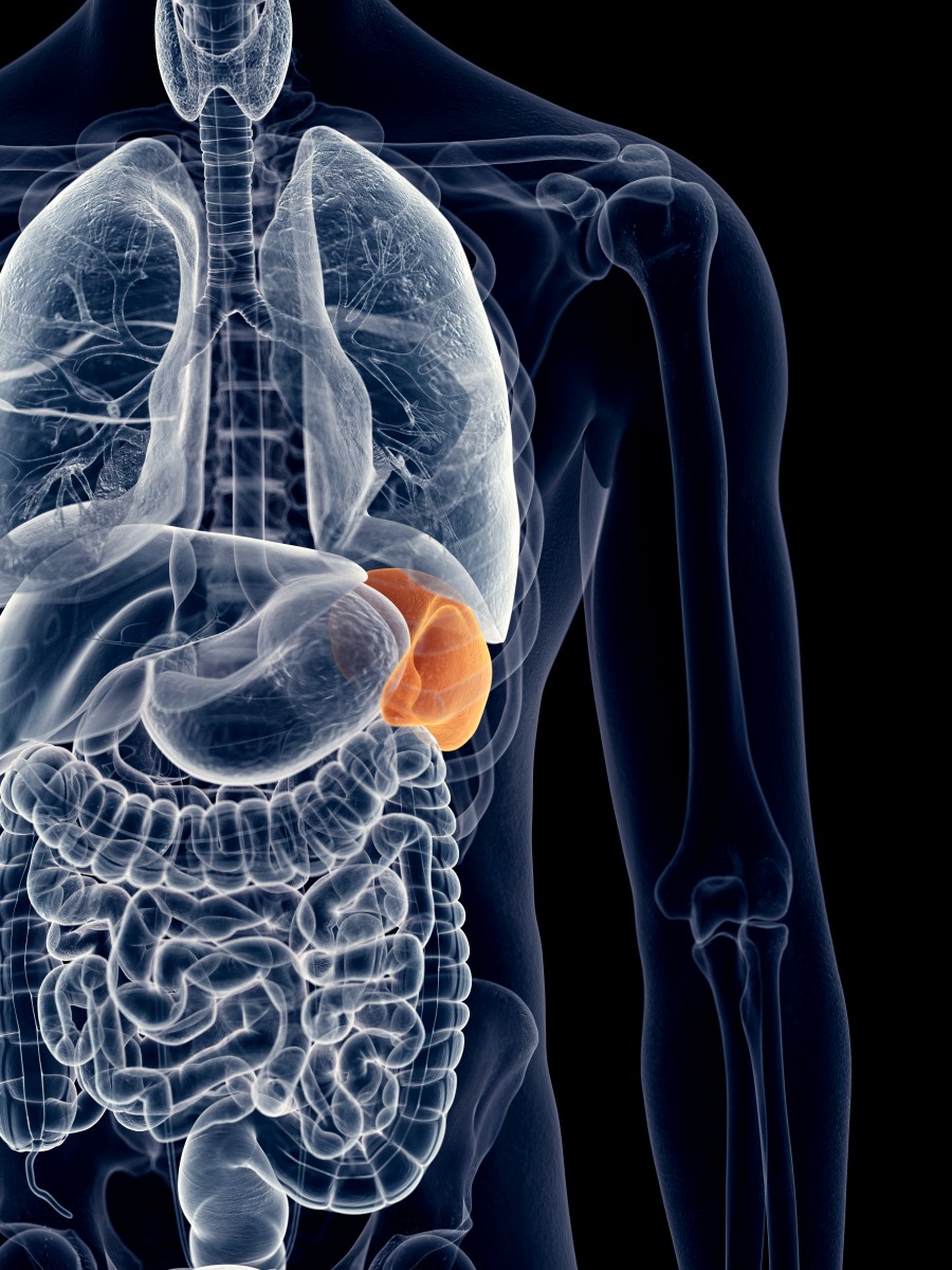
Specifically, Gaucher disease patients’ median stiffness of the spleen was measured at 35KPa and 22KPa, by TE and SWE, respectively. In contrast, these values are different from those exhibited by patients with liver cirrhosis (45KPa and 34.5KPa) and healthy patients (16.95 KPa and 17.5 KPa).
Looking at liver stiffness, researchers observed that Gaucher disease patients showed values of 7.1 KPa and 7 KPa, either by transient and 2D shear wave elastography, respectively. These values were slightly higher when compared to healthy controls. However, they were much lower when compared to cirrhotic patients (medians of 24.2 KPa and 21 KPa).
These results suggest that transient and shear wave elastography are potential noninvasive methods to differentiate Gaucher disease patients from other patients presenting enlargement of the spleen (also known as splenomegaly).