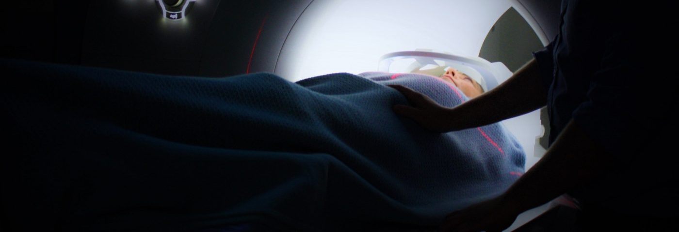
Using magnetic resonance imaging (MRI) to assess water- and fat-associated tissue changes may be used to identify and monitor bone damage in young people with Gaucher disease, researchers from Egypt suggest.
The study, “Multi-parametric MR imaging using apparent diffusion coefficient and fat fraction in quantification of bone marrow in pediatrics with Gaucher disease,” was published in the journal Clinical Imaging.
Among the several organs affected by Gaucher disease, bone lesions — which can affect the spine, pelvis, and long bones of the legs and arms — are the most debilitating.
They are a result of bone marrow infiltration by Gaucher cells, a type of immune cell that shows Gaucher’s characteristic accumulation of specific fat molecules.
This infiltration leads to mechanical obstruction and reduced blood flow, causing cell death and bleeding in the bone. Thus, its early detection and monitoring is important to halt disease progression through enzyme replacement therapy (ERT) and to evaluate treatment response, respectively.
Bone damage can be assessed through various imaging techniques, including MRI. This technique is used to assess the level of bone infiltration and identify lesions and other complications.
But detecting bone lesions through a standard MRI in pediatric patients is challenging due to the small volume of bone marrow and its structural variety during this age period.
However, multiparametric MRI, or the combination of standard anatomical images with functional techniques and parameters, has the potential to be a better method to identify bone damage.
Researchers in Mansoura, Egypt, evaluated whether a multiparametric MRI using two quantitative imaging parameters — fat fraction and apparent diffusion co-efficient (ADC) — could identity and quantify bone marrow damage in pediatric Gaucher patients.
The fat fraction, or the proportion of fat vs. water, usually decreases in Gaucher patients due to replacement of normal rich fat cells with Gaucher cells. The ADC, which assesses the number of cells through the molecular motion of water molecules, is lower in abnormal tissues with a high infiltration of cells.
The study included 29 patients (22 boys and seven girls; mean age of 8) with Gaucher disease type 1, and 13 age- and gender-matched healthy youngsters (mean age 9). Thirteen patients were already being treated with Cerezyme (imiglucerase), an ERT.
The multiparametric MRI with fat fraction and ADC was conducted in the bone marrow of the lumbar spine, or lower back, in all participants.
Both fat fraction and ADC were significantly lower in Gaucher patients than in healthy children, and patients treated with ERT had significantly higher values of fat fraction and ADC than untreated patients.
The team also found a significant association between the values of fat fraction and ADC of the lumbar spinal bone marrow.
The results are consistent with the infiltration of Gaucher cells in the bone marrow of Gaucher patients, and with an effective infiltration decrease with ERT.
These findings suggest that these two quantitative imaging parameters within multiparametric MRI could be used to identify and monitor bone damage in pediatric Gaucher patients.
“Multi-parametric MR imaging using ADC and [fat fraction] are quantitative imaging parameters that can be used for detection and quantification of the bone marrow involvement in pediatric patients with Gaucher disease,” researchers wrote.
The researchers noted, however, that additional studies are necessary to confirm these results and to assess a potential correlation of the fat fraction and ADC of the bone marrow with the different types of Gaucher disease.


