Children with Type 1 Gaucher Disease Show Changes in Motor and Sensory Brain Networks, Study Finds
Written by |
Motor and sensory-related brain networks do not work efficiently in children with type 1 Gaucher disease (GD), but this disrupted performance appears to remain stable over time, researchers report.
Their findings were published in the study, “Altered brain functional network in children with type 1 Gaucher disease: a longitudinal graph theory-based study,” in Neuroradiology.
In type 1 GD, which is the nonneuropathic subtype that is the most common and least serious form of the inherited disorder, the absence of neurological symptoms is considered mandatory for a diagnosis.
However, some studies suggest that neurological abnormalities could be involved in type 1 GD. In fact, researchers have found that some GD patients might develop Parkinson’s disease-like symptoms, including muscle rigidity, resting tremor, and slowed, affected movements.
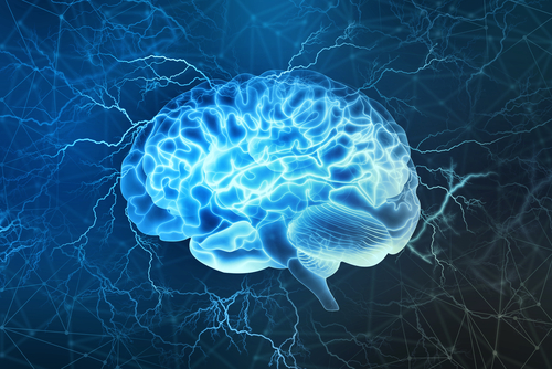
The brain is a complex network of anatomically separate clusters of specialized neurons and functional links between these groups.
“Regions functionally related during a cognitive task tend to be temporally correlated at rest,” the authors wrote.
In addition, structural abnormalities may disrupt the efficiency of communication within brain networks. Researchers at Beijing Children’s Hospital have reported microstructural alterations in the brain’s white matter in children with type 1 GD, but they did not assess the efficiency of the brain functional network.
For this study, the same research team in China set out to examine the efficiency of the whole brain functional network in children with type 1 GD.
They recruited 22 children with type 1 GD and the same number of sex- and age-matched healthy individuals. Participants underwent resting-state functional magnetic resonance imaging (rs-fMRI) procedures twice — at the beginning of the study and at a follow-up 4.6 years later.
The rs-fMRI examination is similar to conventional functional magnetic resonance imaging but does not require subjects to perform a task or respond to stimuli. Patients just lie in the scanner for five to 10 minutes in an awake state while whole-brain blood oxygenation level-dependent (BOLD) data is collected. This data enables investigators to map the brain’s functional connectivity.
Researchers then performed a graph theoretical analysis to evaluate overall brain network properties.
Networks can be represented by graphs, which are sets of vertices (or nodes, represented by filled circles) and corresponding sets of edges (or connections, drawn as lines). The existence of an edge between two vertices indicates the presence of some kind of interaction or connection between the vertices, the meaning of which depends on what the graph is modeling.
Results showed that in comparison with the control group, children with type 1 GD had a decreased efficiency in neuronal processing carried out among functionally-related brain areas arranged within “modules.”
In brain science, modules refer to “communities,” characterized by high density of connectivity among members of the same community plus low density of connections between members of different modules.
The balance between neuronal processing and communication between “communities,” plus the integration of the distributed information, was also compromised in children with type 1 GD.
Researchers also reported that, compared with the control group, the functioning of motor- and sensory-related networks in type 1 GD patients was compromised due to fewer neuronal paths within the net and lower information transfer efficiency from one brain region to another within the same network.
More specifically, the affected motor-related processing region was the right precentral gyrus, which plays a role in motor action preparation and control. The affected sensory-related processing region was the postcentral gyrus, which is involved in pain recognition and tactile discrimination.
Almost five years after the first rs-fMRI, no significant changes were found in the global and regional brain network functioning of the GD group.
“Our study provides new evidence of brain changes to further confirm the concept of the existence of [a diverse collection of disease characteristics] in children with type 1 GD and facilitates the understanding of its neuropathological mechanism. However, our results are preliminary and require further investigation,” the investigators concluded.