Rare Case of Gaucher Disease with Lung Damage Seen in Young Child
Written by |

A case report by researchers at Chacha Nehru Bal Chikitsalya Hospital in India described a child with a rare type of lung involvement in Gaucher disease. The study underscored that lung damage in Gaucher patients might be irreversible, highlighting the importance of an early diagnosis.
The study, “Lung lysed: A case of Gaucher disease with pulmonary involvement,” was published in the journal Lung India.
Researchers described a 6-year-old girl who had a firm lump on the left side of her belly. The lump had been growing for three years and now distorted the shape of her body. The child also had a history of frequent episodes of cough and cold, breathlessness, poor weight gain, and showed signs of a mild delay in her ability to walk and move.
A physical exam revealed that her liver and spleen were enlarged, and doctors noted she was breathing at an abnormally fast rate. Sounds during breathing also indicated airway disease, the doctors reported.
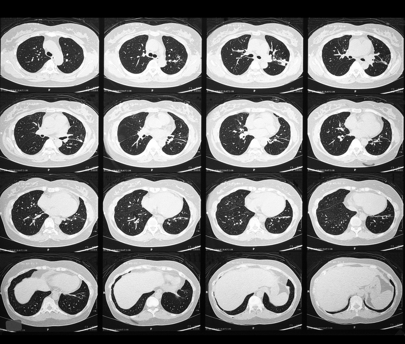
A lung X-ray was taken, and high-resolution computed tomography (HRCT) scans of the chest showed areas with a hazy appearance, referred to as ‘ground glass opacities.’ It was also evident that connective tissue between lung areas, known as lobules, was thicker than normal, a feature doctors call ‘crazy paving appearance.’ This thickening was seen both in the smaller and larger lobules.
An analysis of the bone marrow found cells resembling what is known as ‘Gaucher cells’ — cells that look like crumbled tissue paper. This finding led the doctors to test the girl’s levels of β-glucocerebrosidase, the enzyme whose malfunction causes Gaucher disease, which were low. In addition, levels of the enzyme chitotriosidase were increased 1,000-fold — a typical feature found in most Gaucher patients.
The enzyme measures, along with the clinical findings, led the medical team to set a diagnosis of Gaucher disease, Type III, typically affecting children.
Lung disease is present in more severe forms of Gaucher, and the type of lung damage seen in the girl was unusual since it affected both the connective tissue, and the lung alveoli — the tiny sacs in the lung where oxygen can enter the blood. The damage was also seen only in parts of the lungs — a combination of features that has been described only once before.


